Arterys is a Rosenman Innovator company. From Fierce Biotech
Analyzing the results of an MRI scan can be a complex, time-consuming process for physicians. Outsourcing much of the work to artificial intelligence, however, can slash that time and effort by about a third, according to AI developer Arterys.
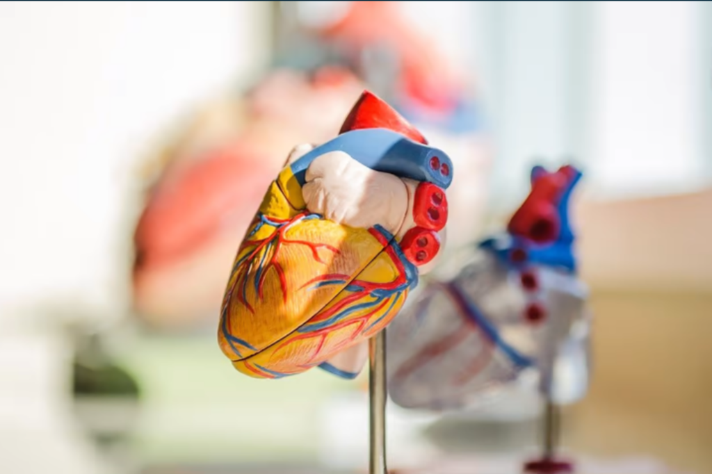
The San Francisco, California-based medical startup has built a handful of tools that use deep learning and other forms of AI to analyze a range of imaging data, including lung CT scans, chest X-rays and both brain and heart MRIs. The last of these is the subject of Arterys’ latest leap forward, which brought in a new FDA clearance—the company’s eighth to date—and added a handful of new features for the Cardio AI solution.
The cloud-based Cardio AI tool uses deep learning algorithms to rapidly take in and assess MRI scans of the heart, producing analyses Arterys says are as accurate as the segmentations performed manually by human experts—but in much less time.
The FDA green light was bestowed upon Arterys’ modules for T1 mapping and T2 quantification. The T1 mapping tool, which also calculates extracellular volume fraction (ECV), looks for variations in the heart muscle’s tissue. Those findings can then be used to diagnose conditions like myocardial inflammation, fibrosis and hypertrophy, while the ECV calculations measure the empty space in the tissue that may also play host to certain myocardial diseases.
T2 quantification, meanwhile, provides another characterization of the tissue of the heart muscle, helping physicians assess the severity of a case of myocardial infarction and detect swelling in patients with the condition.
In addition to the FDA’s signoff of those new features—which will allow them to be made commercially available in the U.S.—Arterys unveiled three other modules for the Cardio AI solution.
The first of these is a research-use-only tool that’s able to quantify ventricular dysfunction, adding overlays to the MRI that depict strain, strain rate and myocardial velocity. Another new addition assesses the volumes of each of the right and left atria, while the third automatically calculates the thickness of the heart’s walls.
Those and the T1 and T2 quantification tools join other features already embedded in the Cardio AI platform that offer 2D and 4D visualizations of the blood flow through the heart, semi-quantitative analyses of how well the muscle is functioning and more.